


HDlive is an extraordinary rendering method generation amazingly realistic images of the human fetus from songraphic data. Through the use of an advance illumination model, HDlive supports shadows, a virtual light source and advanced skin rendering techniques.
This will reset is amazing realism for the fetus images.
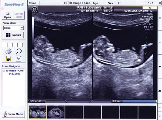
Medical ultrasound imaging utilizes very high frequency sound waves (hence the name “ultra” sound). When these sound waves penetrate the body, they are reflected differently depending on the density of the tissues they traverse. The image seen on the ultrasound monitor depends on the distance between the ultrasound probe and the target tissue as well as on the intensity of the reflected ultrasonic waves thus generated. Such an image is a two dimensional (2-D) image as seen on most ultrasound monitors. This is a simple and safe office examination capable of giving valuable information regarding the pelvic organs such the uterus and ovaries in general, as well as the presence of uterine fibroids, ovarian tumors or cysts, abnormal fluid collection and other abnormalities.In case of pregnancy, ultrasound imaging also provides valuable information regarding the embryo or fetus, its viability, growth, gender and normality. Ultrasound technology progressed in recent years to enable us to use a third dimension – depth, in addition to length and width, to give us a 3D image which enables us to see a three dimensional image of the baby or any organ within the body. With the advent of more powerful computers, we now have the ability to see this 3D image in real time, adding a 4th dimension (time) giving us the opportunity to have an even clearer and more detailed image of babies within the uterus.
The development of these new technologies has not replaced the use of classical 2-D Ultrasonography which is still the basic tool for assessing early fetal growth and development as well as looking for any fetal abnormalities. 4D can be used at a later stage to look at the overall appearance of the fetus.
The three stages of pregnancy can be assessed by 2-D Ultrasound:
1. The first trimester: Starting at 5-6 weeks from the date of the last menstrual period, the presence or absence of a fetal sac within the uterus is documented and any fetal sac outside the uterus) ectopic pregnancy) is carefully looked for. Shortly thereafter, embryonic tissue and heart beat activity are sought, the number of embryos and measurement of the embryo’s size (Crown Rump Length – CRL) determined to help assess the correct age of the pregnancy. By 8-10 weeks the fetal head, body and limbs can be seen and the size of the yolk sac noted. At 11-12 weeks fetal growth and fetal well-being can be further assessed. The thickness of the skin at the back of the embryo’s neck, known as the Nuchal Translucency (NT) is measured in an effort to gauge the risk of trisomy 21 or Down’s syndrome. Cystic hygroma can also be detected, suggesting possible chromosomal fetal anomaly as well.
2. The second trimester of pregnancy is the most important for fetal ultrasound assessment because fetal organs have been formed and their evaluation can be performed with great accuracy
The fetal head and its contents can be seen clearly. Absence of part of the head (anencephaly), small head size (microcephaly), large head size (hydrocephaly) due to excess intracranial fluid, the presence of brain tissue outside the cranium (myelocoele, meningomyelocoele) and other anomalies can be detected at this stage.
The face, neck and nose: Absence of the fetal nose bone is suggestive of Downs’s syndrome. Cleft lip and cleft palate as well as a small jaw (micrognathia) can also be determined.
Gastrointestinal tract: Here we look for the presence and correct position of the stomach, duodenal obstruction, diaphragmatic hernia and intestinal abnormalities. As for the genitourinary tract, kidney and bladder abnormalities and obstruction of the ureters can be detected.
Skeletal system: Length or shortness of the fetus, the number of fingers, defects in the spine, club foot, and absence of limbs (limb reduction) can all be detected.The importance of this examination reflects the accuracy of early fetal diagnosis whose importance is to alert the physician and parents of the unborn child regarding any possible urgent medical or surgical treatment that the child might need right after being born.
3. The third trimester: At this stage we can clearly assess growth of the fetus, its breathing, movement of the body and extremities, and normal or abnormal heart beat. Placental problems such as early separation (abruptio placenta) or abnormal location (placenta praevia) can easily be detected as well as placental function and umbilical cord blood flow via color Doppler examination. Abnormal amount of amniotic fluid can easily be seen – too much (polyhydramnios) or too little (oligohydramnios) being important findings. A tendency to premature delivery could also be suspected by finding shortening or dilatation of the cervix.
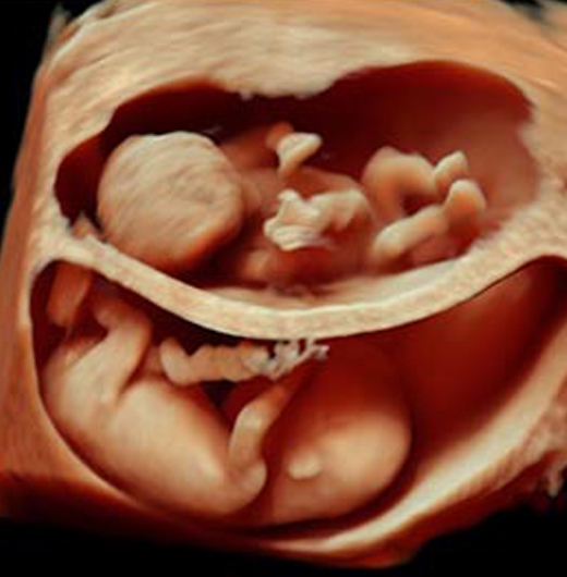
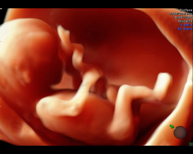
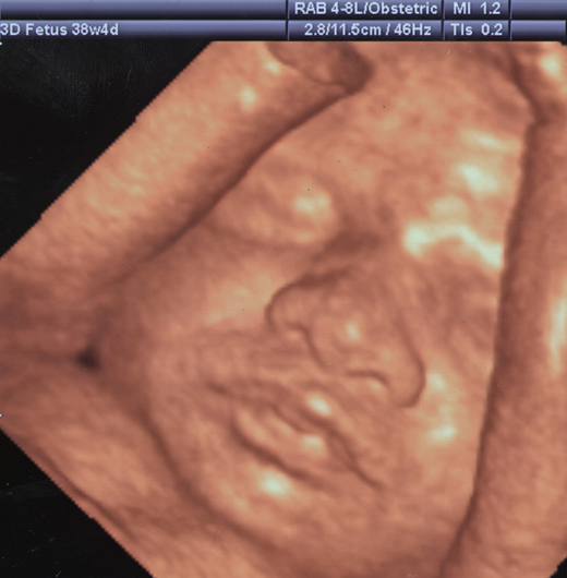
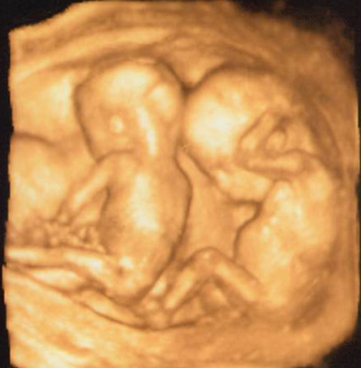
3D Ultrasound allows us to measure the size and appearance of internal organs with great accuracy. The skeletal system can be seen as clearly as by X-Rays. 4D Ultrasound is similar to 3D, but now the images are seen in real time enabling us to visualize the fetal surface without penetrating the body, thus giving the impression of a photographic image of the fetus and showing the external appearance of the face, lips, nose, mouth and ears as well as hands, fingers, toes and genitalia. 4D Ultrasound has given us an insight into the previously hidden aspects of fetal life.
Goals of Ultrasound examination:
The development of 4D Ultrasonography has allowed us to go one step further in our understanding of the details of life inside the womb, the benefits of which are many:
1. The physician can follow up the pregnant mother and her baby at all stages of the pregnancy and in ever greater detail and to interfere at an appropriate time when and if necessary to safeguard the mother’s and baby’s well-being.
2. The early detection of congenital fetal abnormalities, their type and severity which can also help prepare the prospective parents psychologically to recognize and accept the implication of a possible deformity or abnormality, as well as be ready for the cost of treating and bringing up such a child.
3. The social impact on the family of the unborn child seeing live images of their beloved child is a pleasure never to be forgotten. Such images can also be stored digitally and preserved for later viewing by all.
Besides the routine use of Ultrasonography for all pregnant women, the test is particularly valuable for:
- Women over 35 years of age.
- The presence of a child with a congenital anomaly in the family.
- The finding of abnormal laboratory tests in the mother.
- A family history of certain types of hereditary disorders.
- Exposure of the pregnant woman to certain drugs, toxic materials or X-Rays.